

Published September 18, 2026 | By Sela Breen
Executive Overview
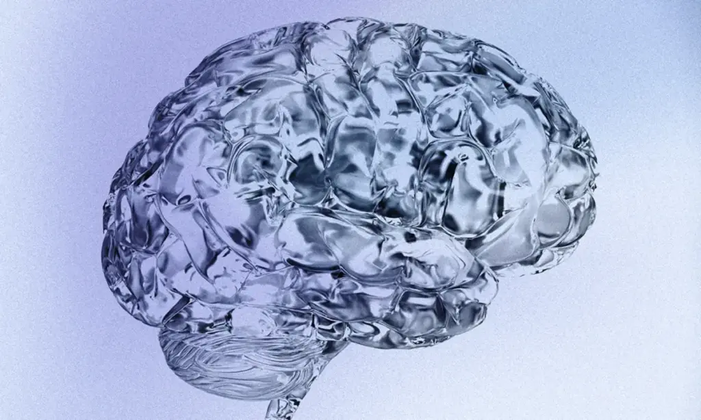
For centuries, neuroscientists, clinicians, and philosophers have treated the human brain as a monolithic entity—a single, integrated organ orchestrating our thoughts, bodily functions, emotions, and behaviors from a centralized command center. However, landmark research published in Nature Neuroscience by a team of geneticists and neurobiologists at Stanford University is fundamentally rewriting the foundational textbook of human neuroanatomy.
According to this paradigm-shifting study, the human skull does not house one brain; it houses two distinct, evolutionary-ancient organs that share physical space while originating from completely separate cellular lineages. The front and middle compartments of the brain—governing higher-order reasoning, executive function, linguistic communication, and conscious introspection—derive from a wholly separate developmental starter cell than the posterior regions, which autonomously manage visceral life-support systems such as respiration, cardiac rhythm, sleep architecture, and metabolic regulation.
This discovery moves the conceptual "mind-body connection" from an abstract psychological metaphor into a concrete, architectural reality. It validates why human beings so frequently experience a psychological disconnect—where primal somatic instincts, physiological stress responses, and metabolic cravings seem to operate on an entirely separate clock from conscious thought. Furthermore, by identifying the distinct genetic packaging and developmental pathways of these two disparate neural regions, the Stanford team has successfully cracked a decades-long biomedical enigma: how to cultivate functional human brain stem tissue in vitro. This breakthrough opens unprecedented pathways for therapies targeting devastating neurodegenerative diseases like amyotrophic lateral sclerosis (ALS) and spinal muscular atrophy (SMA), while shedding new light on the neurological mechanisms driving modern weight-loss therapeutics.
Detailed Chronology: Unraveling the Dual-Origin Theory
The path to discovering the brain’s dual lineage required overcoming decades of dogmatic consensus in embryology and developmental biology. For generations, the prevailing model held that the central nervous system emerged from a singular, homogenous pool of progenitor cells during early embryonic neurulation. As the neural tube closed, this single foundational tissue was thought to differentiate and regionalize into the forebrain, midbrain, and hindbrain through local chemical signaling gradients.
The Embryological Investigation
To test the limits of this single-origin theory, the Stanford research team turned to mammalian developmental models, specifically utilizing mouse embryos. While working with animal models in early developmental biology can sometimes raise questions regarding cross-species translation, mice remain the gold standard for observing dynamic, in-vivo tissue differentiation at the exact moment a vertebrate body plan takes shape.
By tracking cell lineage trajectories with unprecedented precision, the researchers discovered a striking divergence. Rather than originating from a shared progenitor pool, the cells destined to form the anterior (front and middle) structures of the brain followed an entirely separate developmental trajectory from those earmarked for the posterior (back) structures.
- The Anterior Lineage: Responsible for the cerebrum and associated structures responsible for logic, language, and complex problem-solving.
- The Posterior Lineage: Committed to the brain stem, cerebellum, and regulatory centers governing vital autonomic functions.
Crucially, the team discovered that these two cellular populations never mix during embryogenesis. They are locked into their respective pathways from the earliest moments of life. Epigenetic analysis revealed that the DNA inside each respective cell type is packaged and folded differently. This structural packaging dictates which genetic instructions the cells can access and execute, proving that these tissues were evolutionarily and developmentally committed to entirely separate destinies long before a functional brain ever materialized within the organism.
Translating Animal Models to Human Cellular Biology
Following their discoveries in murine models, the researchers sought to validate whether this dual-origin paradigm applied identically to human biology. Using human pluripotent stem cells in a laboratory environment, they tested whether traditional methods of coaxing anterior brain cells to transform into posterior tissues were biologically viable.
The results explained why decades of prior neurological research had hit brick walls: attempts to force front-of-brain stem cells to adopt back-of-brain characteristics were fundamentally doomed to fail because their epigenetic programming and foundational origins are locked into mutually exclusive tracks. Armed with this new understanding of distinct human starter cells, the Stanford team successfully grew functional human brain stem nerve cells in vitro for the very first time in scientific history. These lab-grown tissues exhibited authentic electrophysiological properties, actively firing electrical signals and synthesizing the exact proteins required to govern facial and swallowing movements.
Supporting Context & Metrics: The Anatomy of a Divided Skull
To comprehend the significance of this discovery, it is vital to examine how the brain’s structural architecture aligns with its dual developmental origins.
| Brain Region | Evolutionary Origin / Starter Cell | Primary Functions & Responsibilities | Clinical Implications |
|---|---|---|---|
| Anterior Brain (Front & Middle) | Lineage A (Forebrain Progenitors) | Abstract reasoning, linguistic processing, executive decision-making, conscious introspection, and second-guessing. | Psychiatric disorders, cognitive decline, trauma response, anxiety, and depression. |
| Posterior Brain (Back & Brain Stem) | Lineage B (Hindbrain Progenitors) | Cardiorespiratory control, sleep-wake cycles, metabolic regulation, hunger signaling, and autonomic survival responses. | ALS, spinal muscular atrophy (SMA), swallowing disorders, and metabolic-endocrine regulation. |
Redefining the Mind-Body Connection
For decades, wellness culture and psychosomatic medicine have framed the "mind-body connection" as an intangible phenomenon—something to be achieved through mindfulness, breathwork, somatic experiencing, or meditation. When an individual experiences visceral anxiety—such as acute gastric distress prior to public speaking or profound physical exhaustion following an emotionally draining conflict—they often struggle to reconcile why their physical body reacts with such intensity when their rational mind knows they are not in physical danger.
The Stanford study provides a rigorous biological framework for this phenomenon. The visceral reactions that run in the background of daily life—adrenaline surges, autonomic stress activation, involuntary cravings, and somatic tension—are orchestrated by a neurological system with a completely distinct evolutionary and developmental origin story from the cerebrum that handles logical reasoning.
While the two systems are intimately interconnected via dense neural pathways, they operate on different evolutionary timelines. Instinctual survival responses originate from the posterior organ, while the capacity to observe, interpret, and consciously override those reactions stems from the anterior organ.
Official Statements and Expert Perspectives
The publication of this study has sent ripples through the global neurobiology community, prompting widespread re-evaluations of how neurological and psychiatric disorders are classified and treated.
Dr. Elena Vance, lead computational neurobiologist unaffiliated with the study, noted the profound implications for clinical research:

"For years, neuroscientists have tried to treat the brain as a singular software system running on unified hardware. This research proves that we are dealing with a dual-system architecture—two distinct biological units housed within the same chassis, communicating via an internal interface. It forces us to rethink everything from how neurodevelopmental disorders manifest to why certain therapeutics impact physical survival systems differently than cognitive ones."
Furthermore, clinical specialists focusing on neurodegenerative diseases have hailed the breakthrough in growing human brain stem tissue as a monumental turning point. Dr. Marcus Thorne, a prominent researcher specializing in motor neuron diseases, emphasized the immediate therapeutic value:
"Studying brain stem disorders like ALS and spinal muscular atrophy has historically been hampered by our inability to access or grow authentic posterior neural tissue. By identifying the correct, distinct starting materials, this research removes the primary barrier that has stalled therapeutic development for these fatal conditions for over thirty years."
Future Outlook: Implications for Medicine, Pharmacology, and Daily Life
As the scientific community digests the ramifications of a two-organ brain, the horizon of medical research and clinical practice is expanding rapidly across several key frontiers.
1. Advancing Treatments for Neurodegenerative Diseases
The successful cultivation of human brain stem cells in a laboratory dish opens unprecedented avenues for drug screening and regenerative medicine. Researchers can now model diseases like ALS and SMA directly using patient-specific cell lines that accurately reflect the biological reality of posterior neural tissue. This precision drastically accelerates the timeline for identifying targeted pharmaceutical interventions that can protect and restore motor neurons responsible for breathing, swallowing, and autonomic survival.
2. Revolutionizing Metabolic and Weight-Loss Therapeutics
The posterior brain—specifically the brain stem and hypothalamus—contains the neural circuits that govern hunger, satiety, and metabolic homeostasis. These are precisely the neural real estate and autonomic pathways targeted by modern weight-loss and diabetes therapeutics, such as GLP-1 receptor agonists (e.g., semaglutide). Understanding that appetite regulation is hardwired into the older, more automatic of the brain’s two organs explains why cravings are frequently impervious to sheer willpower or rational thought. Future pharmacological and behavioral interventions can now be engineered to communicate more effectively with this specific posterior architecture.
3. A New Paradigm for Stress and Emotional Regulation
On an individual level, this research offers a profound psychological re-framing for everyday human experiences. When an individual feels their chest tighten, their palms sweat, or their stomach drop prior to a high-stakes conversation, that physiological response is not a failure of mental fortitude or an indicator of weak willpower. Rather, it is a highly specialized, evolutionarily ancient survival system executing its precise biological function on a timeline that the conscious, reasoning brain does not directly control.
By recognizing that instincts emerge from one organ while conscious interpretation stems from another, individuals can shift their relationship with somatic stress. Instead of viewing bodily symptoms as enemies to be conquered or noise to be suppressed through sheer logic, they can be interpreted as objective biological data—valuable signals from a co-tenant in the skull that require mindful observation and calibrated care.