

Executive Overview
In the relentless pursuit of treatments for some of humanity’s most devastating neurological and psychiatric conditions, a team of pioneering researchers has crossed a formidable scientific threshold. Led by Professor Sergiu Pașca at Stanford University, scientists have successfully implanted human brain-like tissue—known as organoids—into the developing brains of newborn laboratory mice. This ethically sensitive, technically audacious breakthrough aims to solve one of modern medicine’s most frustrating bottlenecks: the profound inability of traditional animal models to accurately replicate complex human brain disorders.
For decades, preclinical drug development for psychiatry and neurology has operated under a dark cloud of inefficiency. Psychiatry famously suffers from one of the lowest success rates in clinical trials across all of medicine. Pharmaceutical candidates that perform brilliantly in rodent models routinely fail when translated to human clinical trials. This chasm in efficacy highlights a stark biological reality: mice and humans are separated by millions of years of evolutionary divergence. Rodents simply do not develop many of the sophisticated brain disorders that plague humankind, such as schizophrenia, autism, severe epilepsy, and cerebral palsy.
By utilizing mice essentially as living biological incubators for human neural tissue, the Stanford team has forged a new hybrid model. This technique allows researchers to observe human brain cell development, circuitry formation, and electrophysiological activity in a living, breathing mammalian brain for the first time. While the architecture of these hybrid brains is admittedly unorthodox and structurally complex, the methodology opens a revolutionary window into human neurobiology. Yet, as this research pushes past traditional boundaries, it simultaneously forces the scientific community, ethicists, and the public to confront profound questions regarding animal welfare, cognitive alteration, and the very definition of what it means to blur the lines between species.
Detailed Chronology: How the Breakthrough Was Achieved
The journey toward successfully integrating human neural tissue into a rodent host required a meticulously choreographed sequence of genetic engineering, cellular reprogramming, and neurosurgical precision.
Step 1: Engineering the Rodent Host
To give human cells the best possible chance of survival and integration, the researchers had to circumvent the natural biological barriers posed by a fully formed mouse brain. The Stanford team genetically engineered laboratory mice to develop with a severely compromised cerebral cortex—the outer layer of the brain commonly referred to as "grey matter," which is responsible for higher-level thinking, sensory processing, and memory. By limiting the development of the mouse’s own cortical tissue, researchers created a vital neuroanatomical "vacancy" within the host cranium, preventing the human tissue from being outcompeted or rejected by pre-existing murine neural networks.
Step 2: Reprogramming Human Cells into Organoids
Concurrently, the research team harvested ordinary somatic cells—specifically skin cells—from human donors. Through advanced cellular reprogramming techniques, scientists induced these skin cells to revert into pluripotent stem cells, which were subsequently coaxed into growing and differentiating into three-dimensional structures known as brain organoids.
It is a common misconception that these organoids represent miniature, thinking human brains grown in laboratory glassware. In reality, they are sophisticated, multicellular aggregates of living human neural tissue. They mimic the early developmental stages of the human cerebral cortex, containing various types of neurons and supporting cells. However, they lack sensory inputs, systemic blood supplies, and the overarching structural organization of a natural, whole human brain.
Step 3: Surgical Implantation and Integration
During the early postnatal stages of the engineered mice, the human brain organoids were surgically implanted directly into the depleted cortical regions of the host animals. Once introduced, the human cells displayed a remarkable degree of biological agency. They did not merely sit inertly within the foreign environment; instead, the cells actively divided, migrated, and organized themselves.
Over a period of months, these human neurons extended axons and dendrites, weaving themselves intricately into the existing neural circuitry and spinal cord pathways of the host mice. Scans of these hybrid brains initially revealed a chaotic, structurally irregular layout—what neuroscientist Dr. Ilary Allodi described as looking "a bit messy" compared to the pristine, highly structured layers of a normal mammalian cortex. Nevertheless, biological adaptation prevailed. Within months, the human cells matured, morphologically and functionally adapting to harmonize with the host’s underlying physiological needs.
Step 4: Behavioral Monitoring and Motor Function
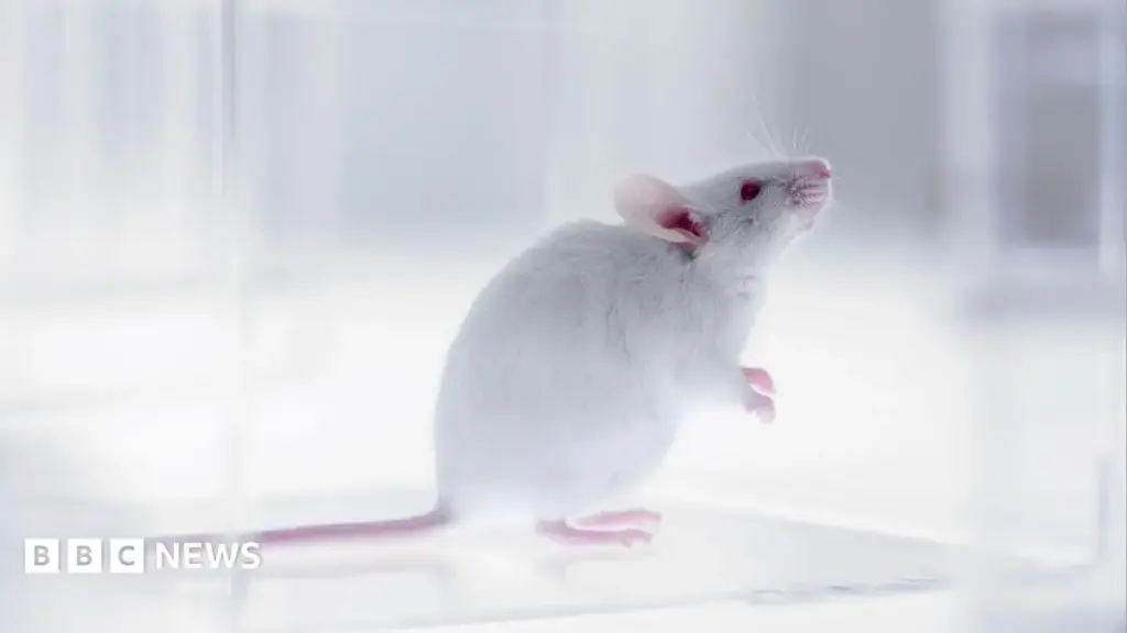
Approximately six months following the surgical procedure, the research team subjected the hybrid mice to a series of baseline behavioral assessments. Placed within a small, tabletop arena, the animals were observed as they navigated their environment.
According to Professor Pașca, the mice performed "largely as [normal] mice did." Crucially, behavioral observation confirmed that the introduction of human neural tissue did not imbue the rodents with any cognitive enhancements or superhuman abilities. Their baseline instincts, movement patterns, and exploratory behaviors remained thoroughly murine, providing vital reassurance during the early stages of this controversial research avenue.
Supporting Context & Metrics: The Crisis in Translational Neuroscience
To truly appreciate the necessity of Professor Pașca’s research, one must examine the staggering failure metrics defining contemporary pharmaceutical research in neurology and psychiatry.
The Clinical Trial Crisis
Clinical trials targeting psychiatric and neurological disorders face a failure rate that outstrips almost every other medical discipline. According to industry analyses, over 90% of candidate drugs entering clinical trials for Alzheimer’s disease, for instance, fail to secure regulatory approval. For psychiatric conditions like major depressive disorder, treatment-resistant depression, and schizophrenia, the pipeline is similarly plagued by high attrition rates.
The core issue lies in the reliance on reductionist models. Historically, neuroscientists have had to rely on a precarious triad of experimental platforms:
- Rodent Models: While genetically tractable, mice possess brains structurally and functionally distinct from humans, particularly in the expansiveness and complexity of the cerebral cortex.
- In Vitro Cell Cultures ("Cells on a Plate"): While useful for observing isolated biochemical reactions, two-dimensional cellular cultures fail to replicate the rich, three-dimensional spatial architecture and synaptic connectivity of living brain tissue.
- Computational Neural Networks: Computer models offer theoretical simulations but lack the chaotic, adaptive, and biochemical reality of organic life.
The Evolutionary Gap
The genetic and physiological gap between mice and humans is vast. Notably, the human brain contains specific neuronal subtypes—such as particular classes of interneurons and projection cells—that are entirely absent in the brains of mice and other rodents. When researchers attempt to study human-specific neurodevelopmental disorders like autism spectrum disorder, Rett syndrome, or severe childhood epilepsy using conventional murine models, they are often looking through a warped lens. The underlying human pathology simply cannot manifest in a rodent brain that lacks the corresponding cellular architecture.
By utilizing the mouse as an incubator, researchers have managed to preserve the "human program"—allowing human-specific neurons to spontaneously emerge, develop, and form functional networks within a living mammalian system. As Dr. Ilary Allodi noted, this provides neuroscientists with an unprecedented bridge across the evolutionary chasm, offering a tangible avenue to study human disease pathogenesis in real time.
Official Statements and Ethical Perspectives
As expected with any scientific endeavor that bridges species boundaries and involves human neural tissue, the Stanford study has ignited intense debate across the global scientific, philosophical, and bioethical communities.
The Scientific Defense
Defending the necessity of the work, Professor Pașca emphasized during a press conference that the ultimate objective is compassionate and urgent: alleviating the immense human suffering caused by untreatable brain disorders.
"Here we have a new model that allows us to actually capture aspects of human brain function in a way that has not been possible before," Pașca stated. He stressed that without capturing the missing information inherent exclusively to human biology, translational medicine will continue to run aground in clinical trials.
Dr. Ilary Allodi, a neuroscientist from St Andrews University who was not involved in the study, praised the technical ingenuity of the work. She highlighted the spontaneous generation of primate- and human-specific cell types within the mouse environment as a watershed moment for the field.
"You’re keeping the human program inside the mouse environment—like the mouse is an incubator," Allodi remarked to the press, validating the methodological shift away from isolated cellular plates toward integrative in vivo hosts.
The Bioethical Horizon
However, the ethical dimensions of integrating human brain tissue into animals are as complex as the biology itself. Dr. Sarah Chan, a reader in bioethics at the University of Edinburgh who was also independent of the study, offered a measured perspective to BBC News.
Dr. Chan confirmed that based on current behavioral metrics, there is currently "no indication that what’s being created here are mice that can think like humans, or a human brain in a mouse body."
Nevertheless, she cautioned that the research serves as a profound philosophical catalyst.
"It prompts us to think about what it might mean when we start changing animal cognition," Dr. Chan noted. "How can we know what it’s like to be one of these mice? And how do we take account of that in the ways that we treat laboratory animals?"
Echoing these cautious notes, Dr. James Ainge, a neuroscientist at St Andrews University, raised practical questions regarding the future utility and ethical ceilings of such models. While acknowledging the technical brilliance of the feat, Ainge suggested that the research might ultimately find itself restricted by "the ethical issues of raising living human brain tissue in a mouse and what that would mean for the experience of the animal."
Future Outlook: Where Do We Go From Here?
The successful implantation of human brain organoids into rodent hosts marks the end of one chapter and the harrowing beginning of another. As the scientific community digests the implications of Pașca’s work, several key trajectories will define the future of this research domain.
1. Regulatory and Ethical Oversight
Given the gravity of altering animal neurobiology, future experiments using human-mouse chimeric models will undoubtedly face heightened regulatory scrutiny. Institutional animal care and use committees (IACUCs), alongside international bioethics panels, will need to establish rigorous new frameworks. These guidelines will likely govern the percentage of human tissue permissible in animal hosts, the monitoring thresholds for cognitive enhancement, and strict limits on the types of neurological conditions modeled. The mice engineered in these studies are currently reared under exceptionally strict welfare protocols; maintaining and expanding these standards will be paramount as techniques become more sophisticated.
2. Specialized Laboratories and Targeted Studies
Due to the immense technical difficulty, ethical sensitivities, and resource-heavy nature of the procedure, this technology is unlikely to become a ubiquitous tool in standard biology labs. Instead, it will likely remain concentrated in a small number of specialized research facilities globally. These laboratories will direct the technology toward a narrow portfolio of intractable conditions—such as severe genetic forms of epilepsy, devastating developmental disorders like cerebral palsy, and complex neurodevelopmental syndromes where traditional models have completely failed.
3. Toward Personalized Medicine and Therapeutic Screening
Looking further ahead, the ultimate promise of human-to-rodent organoid transplantation lies in personalized medicine. Imagine a clinical scenario where skin or blood cells are harvested from a patient suffering from a treatment-resistant neurodevelopmental disorder. Those cells could be reprogrammed into patient-specific brain organoids, implanted into murine incubators, and subsequently utilized to test bespoke pharmacological interventions or gene therapies in vivo before administering them to the human patient.
Conclusion
The Stanford University breakthrough is a testament to human ingenuity driven by the desperate need to conquer neurological disease. By fusing human cellular biology with a mammalian host environment, researchers have peered deeper into the black box of the human brain than ever before. Yet, as science marches into territories previously reserved for science fiction, it carries an obligation to proceed with profound humility, rigorous ethical oversight, and an unwavering respect for the boundaries of consciousness and species integrity. The chasm between human biology and effective treatments has finally been bridged—but traversing it safely will require the utmost vigilance from scientists, ethicists, and society alike.