

Executive Overview
For decades, the intersection of art and science has yielded profound revelations about the natural world, often capturing phenomena that exist far beyond the threshold of human vision. At the forefront of this convergence is the annual Nikon Small World in Motion competition, a global premier platform celebrating the art, skill, and scientific utility of microscopic videography and time-lapse photography. Open to researchers, technicians, and hobbyists alike, the competition serves as a vital bridge between rigorous academic inquiry and public engagement, showcasing the breathtaking beauty hidden within cellular structures, aquatic ecosystems, and microscopic organisms.
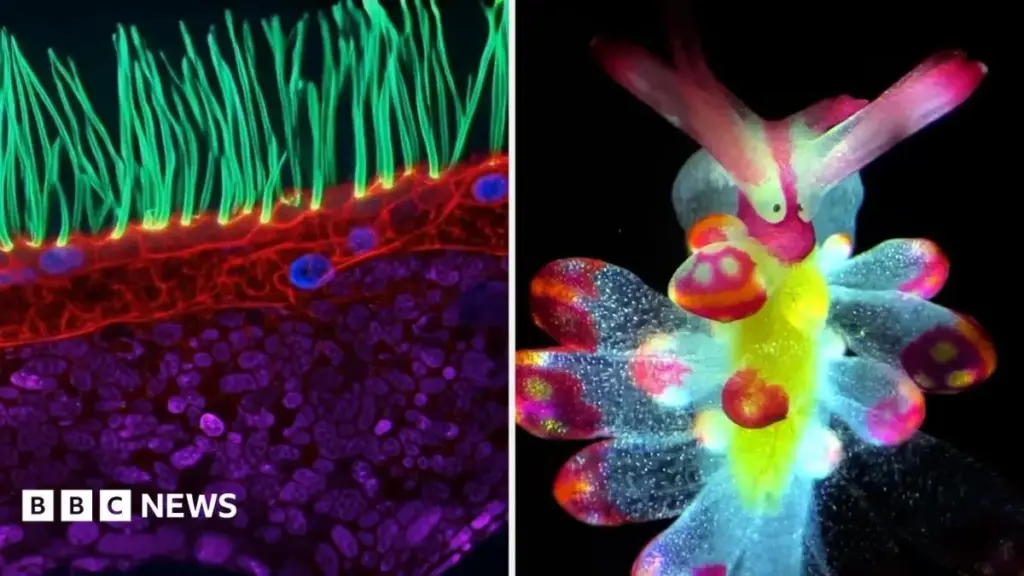
The 2023 edition of the competition has once again disrupted conventional boundaries, culminating in a first-place entry that marries cutting-edge optical engineering with critical pediatric medical research. Submitted by Dr. Ning Xu, a distinguished researcher at Tsinghua University in China, the winning submission offers a mesmerizing, color-enhanced look at the microscopic cilia residing within the human airway of a pediatric patient afflicted with a rare genetic disorder. Magnified one hundred times, the footage is not only an aesthetic triumph of light and motion but also a poignant reminder of the diagnostic and therapeutic potential inherent in advanced optical imaging.
Beyond the grand prize, this year’s competition drew an extraordinary array of submissions from across the globe, featuring everything from predatory encounters between nematodes and single-celled organisms to the ethereal ballet of microscopic jellyfish larvae suspended in micro-droplets. This comprehensive report explores the technical achievements of the winning entries, the sophisticated optical techniques driving modern photomicrography, the broader implications for biomedical research, and the future trajectory of scientific imaging in an increasingly interconnected global research landscape.
Detailed Chronology
The Genesis and Evolution of Microscopic Videography
To understand the significance of the Nikon Small World in Motion competition, one must examine the historical trajectory of photomicrography. Established in 2011 as a companion to the long-running Nikon Small World photomicrography competition (founded in 1975), Small World in Motion was created to acknowledge the rapid evolution of digital imaging sensors and microscope optics. As cameras transitioned from capturing static stills to recording high-frame-rate, high-definition video through microscope objectives, a new medium of scientific storytelling was born.
Over the past decade, the competition has tracked monumental leaps in technology. Early winners primarily showcased basic biological processes, such as cell division or cytoplasmic streaming, captured under standard brightfield or phase-contrast illumination. However, recent years have witnessed a paradigm shift. Entrants now routinely employ advanced modalities—such as structured illumination, light-sheet fluorescence microscopy, and complex laser-scanning techniques—to capture dynamic cellular environments in three and four dimensions.
The 2023 Competition Lifecycle
The journey of the 2023 Nikon Small World in Motion competition began months prior to the public announcement of winners, following an international call for entries. Researchers, independent filmmakers, and university laboratories submitted hundreds of video clips, each representing countless hours of preparation, sample collection, and technological calibration.
The evaluation process is notoriously rigorous. A panel of esteemed judges, comprising prominent science journalists, researchers, and optical engineers, evaluates each submission based on originality, informational content, technical proficiency, and visual impact. The 2023 judging cycle was particularly competitive, with entries spanning diverse fields including developmental biology, neurobiology, marine ecology, and material science.
The Crown Jewel: Dr. Ning Xu’s Winning Submission
At the apex of the 2023 competition stood the submission by Dr. Ning Xu of Tsinghua University. Dr. Xu’s video, magnified 100 times, focuses on the delicate, hair-like structures known as cilia within the respiratory tract of a child diagnosed with a rare genetic disease.
To achieve this remarkable visual feat, Dr. Xu bypassed conventional white-light microscopy. Instead, he utilized a sophisticated series of structured light waves to selectively illuminate different regions of the biological sample. This technique, combined with advanced digital color-mapping, allowed the various components of the cellular landscape to be distinguished with striking clarity. The resulting video displays a synchronized, wave-like beating motion of the cilia—a fundamental biological mechanism responsible for clearing mucus and debris from healthy lungs—while vividly highlighting the structural anomalies and functional impairments characteristic of the patient’s rare genetic pathology.
The Spectrum of Excellence: Runners-Up and Highly Commended Works
While Dr. Xu’s entry claimed the top honor, the competition’s runners-up and highly commended entries demonstrated the sheer breadth of life existing at the micro-scale.
- Second and Third Place Highlights: Among the distinguished runners-up were captivating clips showcasing the predatory dynamics of a roundworm (Caenorhabditis elegans or related nematode species) as it encounters a single-celled organism, capturing the brutal efficiency of micro-predation. Another standout entry featured microscopic jellyfish larvae suspended delicately within micro-droplets of water, revealing the intricate geometry of their developing tentacles and bell structures under polarized light.
- Highly Commended Entries: The pool of highly commended works further expanded the biological tapestry. Researchers submitted mesmerizing footage of bioluminescent algae emitting ethereal blue light in response to fluid shear stress, as well as high-magnification time-lapse clips of tiny sea slugs navigating complex botanical landscapes. Each of these submissions underscored the technical mastery required to maintain sample viability while recording high-resolution video over extended periods.
Supporting Context & Metrics
The Optical Mechanics Behind the Art
Capturing high-resolution video through a microscope is fundamentally different from standard macro or telephoto cinematography. Operators must contend with severe optical limitations, including diffraction limits, spherical and chromatic aberrations, and rapid focal plane degradation.
In the case of Dr. Xu’s winning video, the deployment of complex light waves represents the pinnacle of modern optical engineering. Traditional microscopy often suffers from low contrast when imaging transparent biological tissues. By employing specialized illumination strategies—such as differential interference contrast (DIC) combined with multi-wavelength laser excitation or patterned illumination—researchers can enhance the signal-to-noise ratio, revealing sub-cellular structures that would otherwise remain obscured.
| Metric / Parameter | Typical Microscopic Video Specification | Dr. Ning Xu’s Winning Submission (Estimated/Reported) |
|---|---|---|
| Magnification Level | 10x – 50x (Standard macro/micro) | 100x (High-resolution objective) |
| Primary Subject | General cell cultures / unicellular life | Human pediatric respiratory cilia (rare genetic pathology) |
| Illumination Technique | Brightfield / Phase Contrast | Complex structured light waves & optical sectioning |
| Digital Processing | Basic color balancing | Advanced multi-channel color-mapping and deconvolution |
| Frame Rate | 24 – 60 frames per second | High-speed capture to resolve rapid ciliary beat frequency |
The Biological Significance of Cilia
To fully appreciate the weight of Dr. Xu’s achievement, one must examine the biological role of cilia. Cilia are tiny, microtubule-based protrusions that extend from the surface of many eukaryotic cells. In the human respiratory tract, motile cilia beat in a coordinated, metachronal rhythm, generating a continuous flow of mucus that traps inhaled pathogens, dust, and particulate matter, propelling them upward toward the pharynx where they can be safely expelled or swallowed.
When genetic mutations disrupt the structural or functional integrity of these cellular engines—as seen in conditions like Primary Ciliary Dyskinesia (PCD)—the consequences are severe. Patients suffer from chronic, recurrent respiratory infections, bronchiectasis, and progressive lung damage. By capturing the precise mechanical failure of these cilia in high definition, Dr. Xu’s video provides researchers with invaluable qualitative data that complements traditional genomic and proteomic assays. It transforms an abstract clinical diagnosis into a visible, quantifiable mechanical dysfunction.
Official Statements
The release of the 2023 winners was accompanied by commentary from prominent figures within the scientific imaging community, emphasizing the cultural and scientific value of the competition.
Eric Flem, communications manager for Nikon Instruments, highlighted the evolving role of the competition in bridging scientific discovery and public appreciation:
"Every year, the Nikon Small World in Motion competition reminds us of the incredible dedication and artistry of researchers around the globe. Dr. Ning Xu’s winning video is a masterclass in modern microscopy; it not only captures a mesmerizing biological process with stunning visual clarity, but it also shines a light on the profound impact that rare genetic diseases have on the human body at a cellular level. Through these lenses, we are privileged to witness the unseen complexities of life, captured by individuals who bridge the gap between rigorous scientific investigation and breathtaking visual art."
Reflecting on his achievement, Dr. Ning Xu shared insights into the motivation behind his submission:
"Microscopy is often viewed strictly as a tool for data collection, but within these complex optical patterns lies a profound narrative about health, disease, and the resilience of life. By utilizing advanced light manipulation techniques, our goal was to make the invisible visible—to give researchers and the public a front-row seat to the delicate mechanics of human cellular health. Winning this competition is a tremendous honor, and it validates the collaborative efforts of our team at Tsinghua University in pushing the boundaries of what biomedical imaging can achieve."
Judges panel representatives also noted a distinct trend in the 2023 submissions toward interdisciplinary collaboration. Increasingly, microbiologists are partnering with optical physicists, data scientists, and computational artists to process terabytes of raw video data, utilizing machine learning algorithms to denoise, stabilize, and colorize complex biological footage without compromising scientific accuracy.
Future Outlook
Technological Horizons in Microscopic Imaging
As we look toward the future of scientific cinematography, several emerging technologies promise to redefine what is possible in microscopic observation:
- Super-Resolution Microscopy Integration: Traditional light microscopy is bounded by the Abbe diffraction limit (approximately 200–250 nanometers). However, the continued refinement of super-resolution techniques—such as stimulated emission depletion (STED) microscopy and stochastic optical reconstruction microscopy (STORM)—integrated with high-speed CMOS sensors will soon allow researchers to capture live-action video at the nanometer scale.
- Artificial Intelligence and Machine Learning: The processing of high-frame-rate, multi-dimensional microscopy data generates massive computational bottlenecks. Future workflows will heavily rely on AI-driven image reconstruction algorithms capable of real-time deconvolution, background artifact removal, and predictive frame interpolation, enabling longer and more stable live-cell recordings.
- Optogenetics and Dynamic Manipulation: Beyond passive observation, the integration of light-activated proteins (optogenetics) with advanced optical imaging systems will allow researchers to not only record cellular motion in real time but to physically influence and control cellular processes using patterned light waves during video capture.
Implications for Biomedical Research and Education
The societal impact of competitions like Nikon Small World in Motion extends far beyond academic prestige. By translating complex pathological conditions—such as the pediatric genetic disorder depicted in Dr. Xu’s winning entry—into accessible, visually striking narratives, these initiatives play a critical role in science communication.
In educational settings, high-definition microscopic videography serves as a powerful pedagogical tool, inspiring the next generation of scientists, medical professionals, and optical engineers. Furthermore, as diagnostic imaging continues to merge with advanced videography, the techniques pioneered by competition entrants will likely find direct application in clinical diagnostics, paving the way for real-time microscopic evaluation of live tissue samples during surgical and diagnostic procedures.
Conclusion
The 2023 Nikon Small World in Motion competition has once again demonstrated that the universe hidden beneath our lenses is as vast, complex, and beautiful as the cosmos above. From Dr. Ning Xu’s groundbreaking visualization of human respiratory cilia battling a rare genetic disease to the predatory dances of microscopic nematodes and the delicate poise of jellyfish larvae, these captured moments remind us of the intricate machinery sustaining life on Earth. As optical technology and computational power continue to advance in tandem, the boundary between art and science will continue to blur, opening unprecedented vistas for exploration, discovery, and human understanding.